Vibrational Spectroscopy
Bruker Hyperion FT-IR Spectrometer & Microscope
Bard Hall B56 (Primary Staff: Kevin Silverstein; Secondary Staff: Mark Pfeifer)
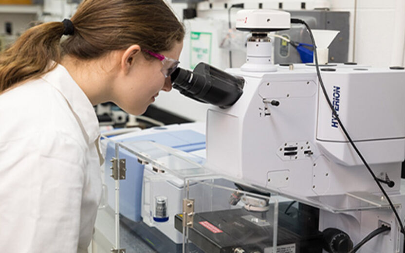
Fourier Transform Infrared Spectroscopy (FTIR) is a spectral analysis technique used to identify chemical bonds. It can be used to investigate existing chemistry, track chemical bonds through a process, or identify unknown materials. Typical samples can be liquids, solids, powders, or even gasses and sample size can be as small as several µl.
The Hyperion is a high performance infrared microscope with transmission, reflection, grazing incidence, and Attenuated Total Reflectance (ATR) acquisition modes. Spectral acquisition area can range from ~250 µm² to as small as ~10 µm². Spectral mapping allows investigation of spatially resolved gradients and distribution of chemical species across a surface.
Bruker Vertex V80V Vacuum FT-IR
Bard Hall SB30 (Primary Staff: Kevin Silverstein; Secondary Staff: Mark Pfeifer)
Fourier Transform Infrared Spectroscopy (FTIR) is a spectral analysis technique used to identify chemical bonds. It can be used to investigate existing chemistry, track chemical bonds through a process, or identify unknown materials. Acquisition of FTIR spectra is most typically performed in 2 configurations: transmission and Attenuated Total Reflectance (ATR). Typical samples can be liquids, solids, powders or even gases. Sample size can be as small as several µL.
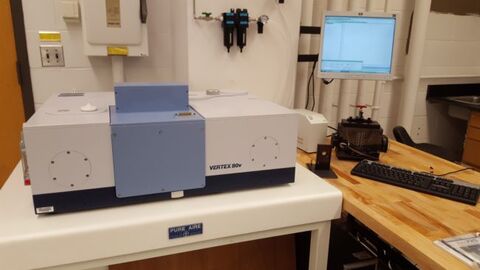
The Vertex V80V vacuum FTIR system provides an evacuated optical bench and sample compartment capable of removing the majority of atmospheric noise. This setup, in combination with a high throughput and sensitive low-temperature MCT detector, provides a very stable measurement platform that is a very effective combination for measuring samples with low signal and/or samples with peaks lying in the water vapor regions.
Renishaw InVia Confocal Raman Microscope
Bard Hall SB30 (Primary Staff: Kevin Silverstein; Secondary Staff: Mark Pfeifer)
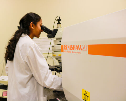
Reflection Raman microscopy enables chemical identification of elements and compounds with a spatial resolution of ~1 µm. Raman shifts within 150 cm-¹ of the excitation frequency can be measured at excitation wavelengths of either 488 nm, 532 nm, or 785 nm, with a spectral resolution of ~1 cm-¹. The 532 nm laser can be used with the ultra-low frequency (ULF) filter to measure Raman shifts as low as 15 cm-1. When the 532 nm laser is used with a 600 l/mm grating and a conventional edge filter, high-speed large-area spatial maps of Raman spectra can be acquired in the Streamline mode.
2D and 3D spatial maps of Raman intensity over areas as large as several square cm can be made using an automated scan stage with any of the lasers–using 3D Volume Mapping of moderately absorbing samples, the Raman intensity can be surveyed to depths of multiple microns into the sample.
WITec Alpha300R Confocal Raman Microscope
Bard Hall SB30 (Primary Staff: Kevin Silverstein; Secondary Staff: Mark Pfeifer)
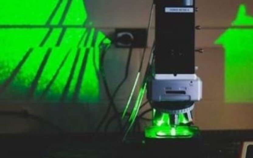
The Alpha300R can be used to investigate existing chemistry, track chemical bonds through a process, investigate crystallinity, image strain fields, and measure photoluminescence. Minimum sample volumes of less than a µL are possible since the analysis is taking place through a microscope. Thin films are very common and in some cases monolayers can be measured.
The Alpha300R incorporates 2 laser excitation sources (532 nm and 785 nm), an advanced confocal microscope, 2 high throughput spectrometers and software capable of powerful multivariate analysis into a system that is built around spectral mapping.