Home⁄ About⁄ News⁄ Studying Diseases with Raman Mapping
Studying Diseases with Raman Mapping
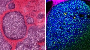
Left, a pathology stain of a ductal carcinoma, showing cells and connective tissue. Right, a Raman mapping of the calcification, which was captured by a technique that detects the distinct vibrational signatures of a biological molecule’s organic and inorganic chemistries.
As described in a recent Cornell Chronicle article and Science Advances paper, the Fischbach and Estroff research groups are studying biomineralization, i.e., how biological organism control the growth of crystals in their tissues. Microcalcifications can capture elements of the tissue microenvironment where they form and in certain cases, can indicate the presence of cancer. This work utilized scanning electron microscopes and the Witec Alpha300R Raman Microscope in the CCMR Shared Instrument Facilities.